Research Paper
Microspore Wall Structure in Selaginella kraussiana (Lycophyta)
John R. Rowley, Marta A. Morbelli and Gamal El-Ghazaly
Published on: June 2002
Page: 115 - 128
DOI: 10.6165/tai.2002.47(2).115
Abstract
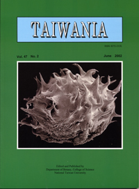
We have examined fresh and macerated microspores of Selaginella kraussiana (Kze.) A. Br. using scanning electron microscopy and chemically fixed spores prepared for transmission electron microscopy. These examinations indicated that the outer spiniferous part of the microspore wall (exospore) is composed of rods that are 70 to 130 nm in diameter and it has numerous conduits of variable size, that pass through the exospore. The structural rods of the exospore are deflected around the conduits. The inner part of the exospore has radial conduits (channels) and it has the same rod-structural units as the outer part. The spines on the outer exospore increase in number and size during development and develop many branches. The outer part of the exospore is separated from the inner part of the exospore by a gap.
中文摘要
以掃描式電子顯微鏡分別觀察新鮮和解離過的卷柏(Selaginella kraussiana)小孢子,另以穿透式電子顯微鏡觀察以化學固定處理後的小孢子。結果顯示小孢子壁外層刺狀部分是由直徑70至130奈米(nm) 的棒狀構造與有許多貫穿小孢子壁大小不一的管線構造組成,棒狀構造繞著管線周圍偏斜著生。小孢子壁的內層的管線構造則呈放射狀,並有與小孢子壁外層相似的棒狀構造。小孢子壁外層的刺狀構造的數目與大小會隨小孢子的發育而增加,形成分支狀。小孢子壁外層與內層是分離的,二者間有一空腔存在。
Keyword: Exospore units, Lycophyta, Microspores, Selaginella, Ultrastructure.


